
Treatment for recurrence
Treatment for recurrence of prostate tumour after irradiation using IRE and HIFU
Radiation therapy is a guideline-compliant and commonly used first-line treatment for prostate cancer. However, similar to other procedures, in 15–20% of cases, cancer may recur within the first few years.
It is usually detected by a PSA rise and a biopsy, and possibly also by an MRI scan or a PSMA-PET scan. PSMA stands for "prostate-specific membrane antigen" and PET for "positron emission tomography".
Since complete removal of the prostate after radiation is difficult and often results in serious complications and a second radiation treatment is generally not possible in such cases, patients are often left with hormone treatment, which has many side effects, in order to at least slowdown tumour growth. As a rule, the effectiveness of hormone treatment diminishes after two to three years, so that at best even more side-effect-laden chemotherapy can then prevent anything worse, at least temporarily.
Recurrence treatment
The Heidelberg Clinic for Prostate Therapy has been successfully using the HIFU procedure for these cases for years. It is one of the few centres that has mastered and regularly uses this procedure, which is now established in the guidelines for such cases. After precise localization diagnostics and histopathological confirmation, the recurrent tumour tissue can be destroyed with pinpoint accuracy, without any further side effects for the patient. Subsequent hormone therapy can be dispensed with in the vast majority of cases.
The situation of a recurrence after brachytherapy is even more problematic. In brachytherapy, radioactive particles remain in the prostate. These represent a contraindication to HIFU therapy. In these cases, IRE treatment can be used to destroy these tumours safely and with few side effects.

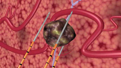
Image: Prostate Therapy Clinic / AngioDynamics.

Photo: Clinic for Prostate Therapy / M. Boeckh